
Technologies
Infection & Immunity imaging
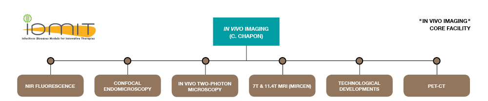
The “Infection &Immunity imaging” core facility constitutes a unique complex combining in vivo imaging and confined facilities for studies of experimentally infected or treated animals.
The imaging suite is accessible for programs of multimodal in vivo molecular imaging using NHP models of human infectious diseases. The suite is located at the CEA Research Center of Fontenay-aux-Roses in facilities suitable for the confinement of class 2 (BSL2) and 3 (BSL3) pathogens if necessary.
EQUIPMENTS/TECHNOLOGIES
- Open imaging system for in vivo fluorescence (FluobeamTM, Fluoptics, near infrared fluorescence with FB700 and FB500) in BSL3 conditions
- In vivo fiber-based confocal microscopy (Cellvizio® DualBand Mauna Kea Technologies) in BSL3 conditions
- Ex vivo confocal fast laser scanning (Nikon®) in BSL3 conditions
- In vivo two-photon microscopy adapted for large animals (Leica®) in BSL2 conditions
- PET-CT suite
AVAILABLE SOFTWARE
- Volocity® software (IRCM, CEA-Fontenay-aux-Roses)
THE MISSIONS OF IN VIVO IMAGING CORE INCLUDE
- Dynamic visualizations of pathogen transmission and dissemination
- Pathogen detection in specific tissue or in NHP whole body
- Dynamic visualizations of host responses
- Monitoring of treatments and prevention strategies
-Biodistribution of drugs, biotherapeutics, antigens and vectors
-Impact on pathogen transmission, dissemination and persistence
-Impact of drugs, biotherapeutics, antigens and vectors biodistribution
-Real-time monitoring of treatments and host responses - Refinement of the use of non-human primate models
-Reduction of invasive approaches
-Limitation of the number of animals
- Monitoring of treatments and prevention strategies
In vivo optical imaging
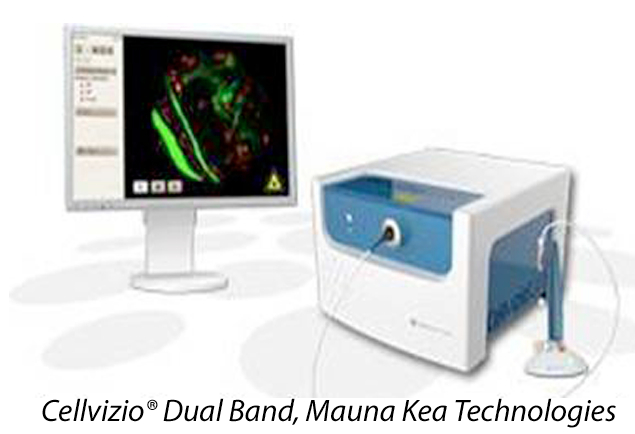
CONFOCAL ENDOMICROSCOPY:
IN VIVO MOLECULAR IMAGING
AT A CELLULAR RESOLUTION
To monitor immune cells at a cellular resolution, in real time.

OPEN IMAGING SYSTEMS FOR IN VIVO FLUORESCENCE IMAGING
To study dynamics of the spread of pathogenic agents/treatments/vaccine vectors in the body.
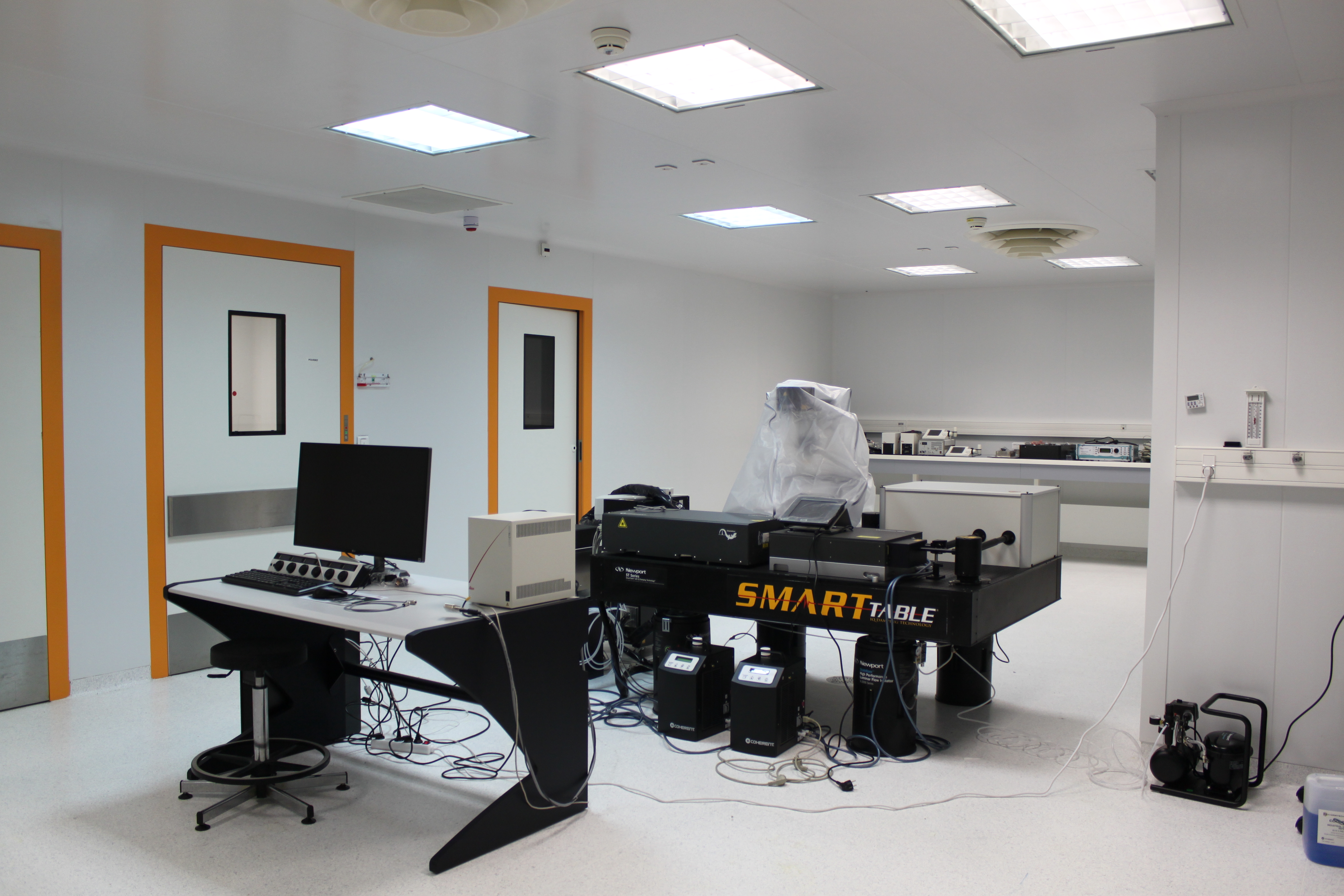
TWO-PHOTON
MICROSCOPY (Leica®)
To explore immune cell interactions with a higher depth penetration in a native environment at a single-cell resolution.
Co-financed by “Union européenne en Ile-de-France avec le Fonds européen de développement régional” and “Contrat de Projets État-Régions 2015-2020 de la Région Ile-de-France“
Ex vivo optical imaging
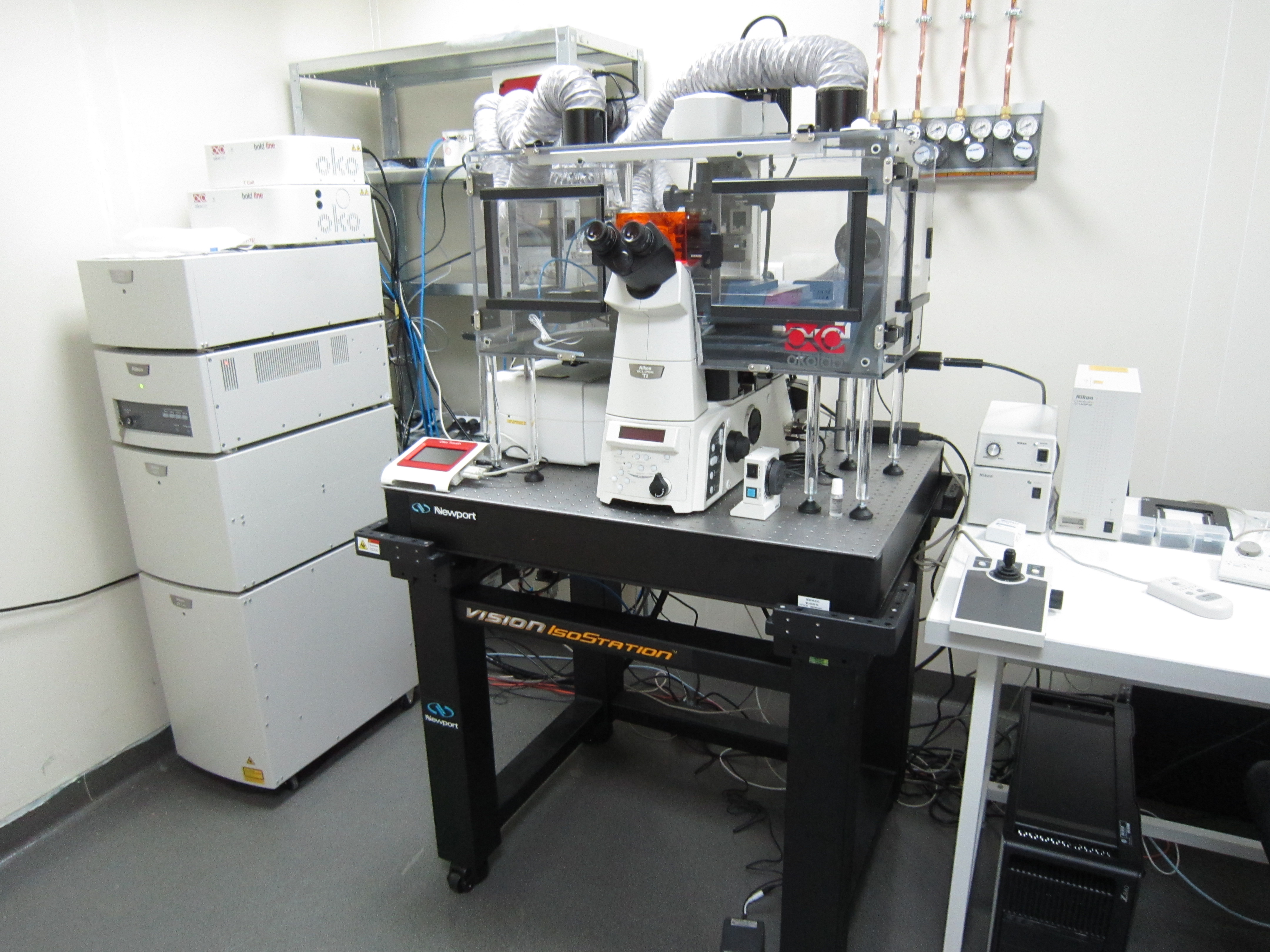
EX VIVO CONFOCAL FAST LASER
SCANNING (NIKON®)
To investigate immune cell behavior on
explants in the context of host response to
pathogens, treatments or vaccination in BSL3 conditions
Co-financed by “Union européenne en
Ile-de-France avec le Fonds européen
de développement régional” and
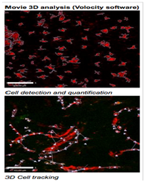
3D TIME LAPSE MICROSCOPY
ON SKIN EXPLANTS
for epidermal APC detection and
quantification, and 3D cell tracking for
analysis of cell velocity and displacement
(using Volocity® software) (B. Todorova)
In vivo PET-CT imaging

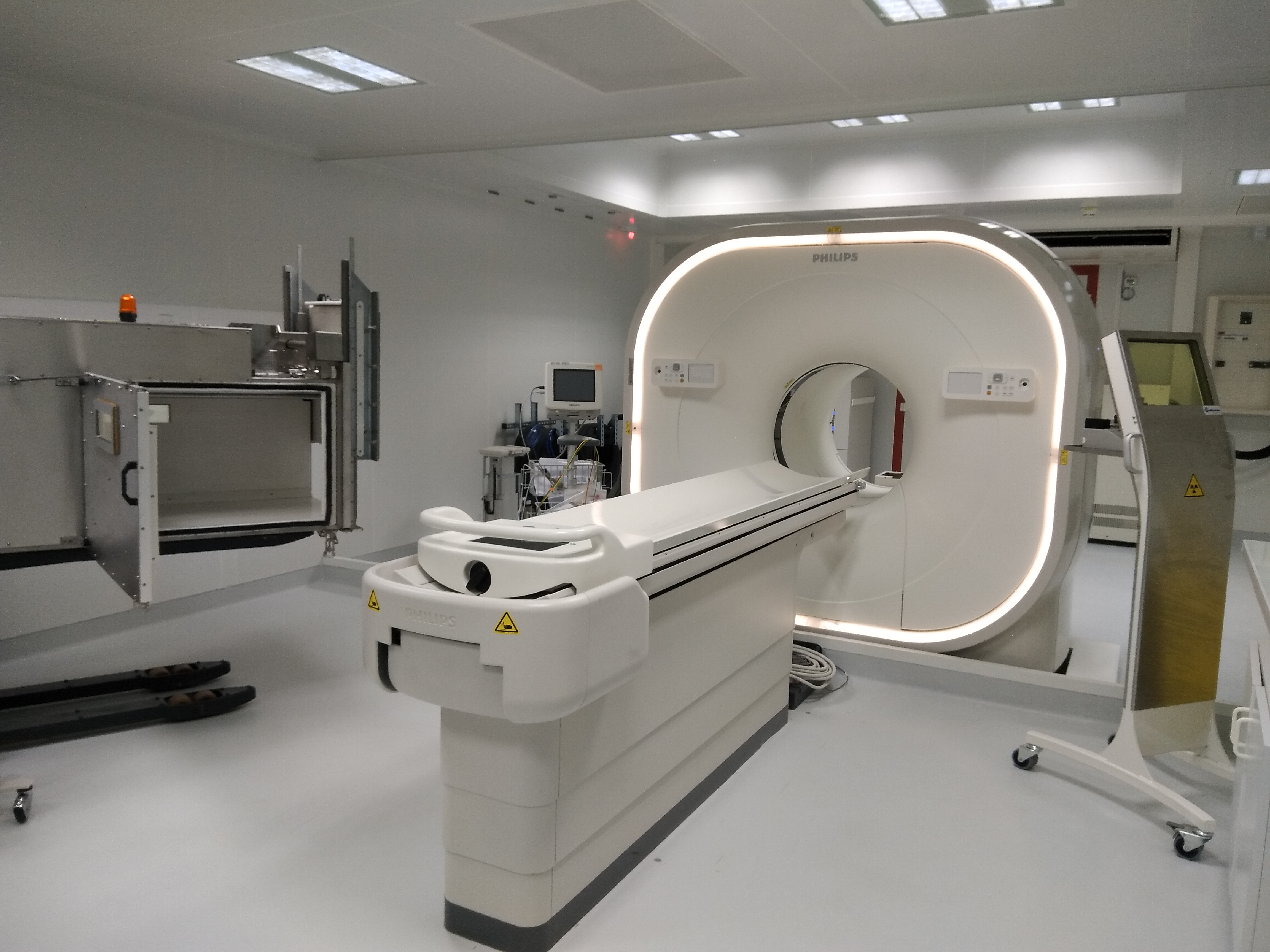
PET-CT (Vereos, Philips)
Co-financed by “Bettencourt-Schueller Fondation” and “Contrat de Projets État-Régions 2015-2020 de la Région Ile-de-France“
To study the whole body drug distribution and the impact of treatments. To identify and follow pathogen sanctuary evolution.
The Vereos digital PET-CT system is more precise, more sensitive and needs shorter time of imaging (Comparison PET-CT).
IDMIT PET-CT system is located in a specific facility and accompanied of different equipment (PET-CT facilities).